In this section we shall explore the bones of the skull and the associated structures:
- The bones of the skull
- The skull base foramina
Once you have reviewed the resources below TEST YOURSELF using our interactive skull base foramina quiz!
THE BONES OF THE SKULL
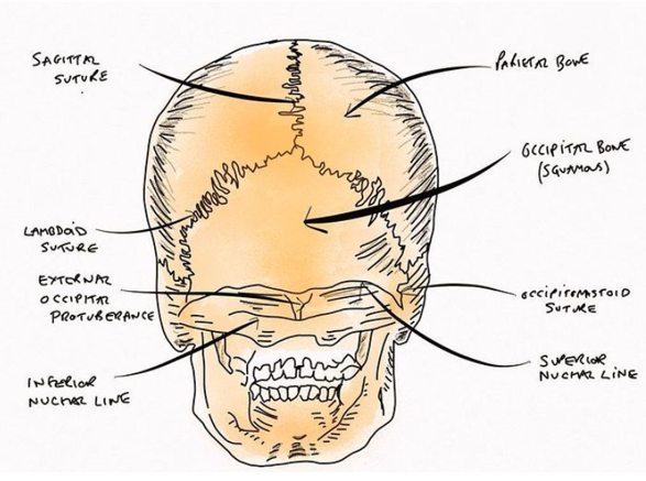
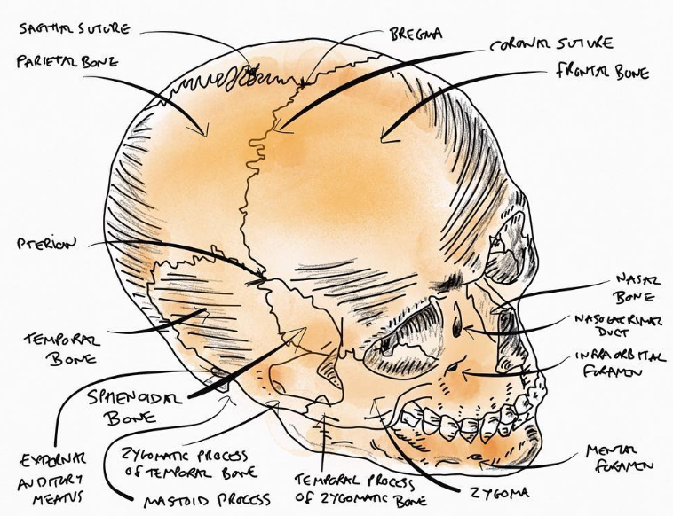
Click on the images above to see 3 different views of the skull bones.
The skull is comprised of a number of bones joined by sutures (fibrous joints). Its purpose is to protect the brain and support the face.
The bones of the skull can be divided into those of the cranium and those of the face
- The face: zygomatic, lacrimal, nasal, palatine, maxilla, vomer and mandible bones and the nasal conchae
- The cranium can be further divided into the cranial roof (calvarium) and the cranial base.
- Cranial roof: frontal, occipital and parietal bones
- Cranial base: frontal, sphenoid, ethmoid, occipital, parietal and temporal bones
THE SUTURES OF THE SKULL
The sutures which join the bones of the skull begin to fuse at the age of 2. Prior to this age, incompletely fused sutures in the skull give rise to the fontanelles- membranous gaps between the bones.
The 2 main fontanelles are the frontal fontanelle and the occipital fontanelle.
- The occipital fontanelle typically closes between 1 and 2 months of age
- The frontal fontanelle typically closes 9 and 18 months of age
Clinical clue 1: The significance of the fontanelle in paediatric examinations
Gentle palpation of the anterior fontanelle can form part of the clinical examination of neonates and infants. The fontanelle should feel firm and flat.
- A sunken fontanelle may indicate dehydration
- A bulging fontanelle may indicate raised intra-cranial pressure, this can have a number of causes:
- Hydrocephalus
- Meningitis or encephalitis
- Trauma
- Hypoxic-ischaemic injury
- Intracranial haemorrhage
Clinical clue 2: Craniosynostosis
Typically the sutures in an infants head will begin to fuse at the age of 2 years. Craniosynostosis is a condition where one or more of the sutures fuse early. This changes the way in which the skull grows. In some cases it grows in a way which still allows sufficient space for the growing brain however, in others it does not.
In cases where the growing brain does not have enough space it will result in raised intracranial pressure. Signs and symptoms may include:
- Visual impairment
- Sleep impairment
- Eating difficulties
- Impairment of cognitive and neurodevelopment function
Craniosynostosis can occur as a part of a syndrome or in isolation.
Craniosynostosis is treated with surgery
Check out our video below for more detail on the bones of the skull:
THE SKULL BASE FORAMINA
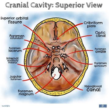
This diagrams demonstrates the foramina of the skull, it is important to know what structures pass through each.
First we shall cover the cranial nerves:
- CN I through the cribriform plate
- CN II and ophthalmic artery through the optic canal
- CN III, IV, V1 and VI through the superior orbital fissure
- CN V2 through foramen rotundum
- CN V3 and lesser petrosal nerve through foramen ovale
- CN VII and VIII go through the internal auditory meatus
- CN IX, X and XI go through the jugular foramen
- CN XII goes through the hypoglossal canal
In addition to the cranial nerves, a number of other structures pass through the foramina of the skull:
- The middle meningeal artery through foramen spinosum
- The spinal cord becoming the medulla oblongata, vertebral arteries and the spinal roots of CN XI through foramen magnum
- The origin of the internal jugular vein is the jugular foramen
And finally it is important to remember that the foramen lacerum is filled by cartilage (don’t get caught out, it is a red herring!)
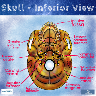
This diagram demonstrates the foramina from the inferior view of the skull. There are a few you can’t see from a superior view:
- The incisive fossa and the greater & lesser palatine foramina, which are associated with the hard palate
- The stylomastoid foramen through which the motor branches of CNVII pass.
- The carotid canal which allows for the internal carotid artery to enter the skull, from here it passes into the carotid canal, then makes its way into the skull by appearing at foramen lacerum.
- Important note here - it does not pass through foramen lacerum!
For more information on the skull base foramina check out our video below: